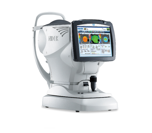
 Computerized Refraction
An automatic refractometer provides an objective measurement of the refractive errors and the prescription for glasses or contact lenses.
-
 ARGOS
ARGOS
With ARGOS we are taking precision and customization to the next level. This advanced biometric system allows us to obtain detailed and accurate eye measurements, which helps to achieve a more precise surgical planning.
-
 Chronos
Chronos offers binocular autorefraction, keratometry measurements and visual acuity through subjective testing. This state-of-the-art technology allows for an accurate assessment of eye health.
 Biometry with IOL Master 700
This test uses the SWEPT Source OCT technology to study the dioptric (corrective) power of the intraocular lens to be implanted in patients who will undergo cataract surgery.
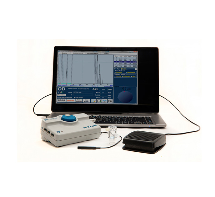 Ocular Biometry by Immersion
This technique provides measurable and reliable data for the correct calculation of the intraocular lens to be implanted in cases where the cataract is so dense it prevents measurement with the IOL Master 700.
 Humphrey Field Analyzer 3 (HFA3) Visual Field Test
The most advanced version of visual field testing, offering faster, more comfortable, and more precise examinations. It allows for the early detection of visual field damage and provides reliable follow-up in glaucoma and other visual conditions.
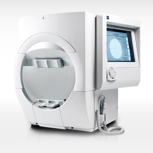 Computerized Humphrey Perimetry
Used to assess the alterations of the visual field, it is essential in cases of glaucoma, since the progressive loss of nerve fibers of the optic nerve results in the loss of certain areas of the field of vision. It is also required in many neuro-ophthalmological diseases.
The aim of visual field exploration is to discover these "blind" areas, locate them and measure their extent.
 ZEISS Clarus 700
It is a complete ultra-widefield retinograph to capture true color images, with unmatched quality and extensive integration of modalities, including fluorescein angiography.
 Endothelial Specular Microscopy
It is a non-invasive technique for the study of the corneal endothelium –the innermost layer of the cornea, responsible for its transparency– which measures the density, size and shape of these cells.
-
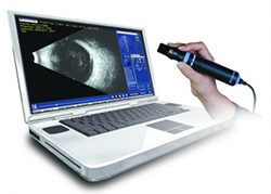 A/B Ultrasound
An imaging test used to diagnose intraocular pathologies of the posterior segment of the eye and the orbit. It is important for the diagnosis and monitoring of some types of uveitis (inflammation of the vascular layer of the eye), intraocular hemorrhages, studies of retinal detachment, foreign bodies in the eye, tumors, ocular trauma, among others.
 Ultrasound Biomicroscopy (UBM)
It allows for an evaluation and measurement of the structures of the anterior segment of the eye through high resolution images, obtained by ultrasound. It is used in cases of hemorrhages and tumors of the anterior segment; subluxated intraocular lenses; traumatic damage to the crystalline; some types of glaucoma; etc.
 OPD Scan III / Corneal Topography and Aberrometry / Pupillometry
A corneal analyzer that combines advanced technologies, including an infrared laser which allows the eye to be fully evaluated, providing data such as autorefraction, corneal topography, keratometry, pupillometry and aberrometry studies for cataract and refractive surgeries.
-
 Pentacam AXL Wave (Ocular Tomography)
The new Pentacam AXL Wave is the first device to combine five parameters: Scheimpflug tomography with axial length, total wavefront, refraction, and retroillumination. Pentacam technology measures, displays and analyzes the anterior eye segment, and is non-contact and tear-film-independent.
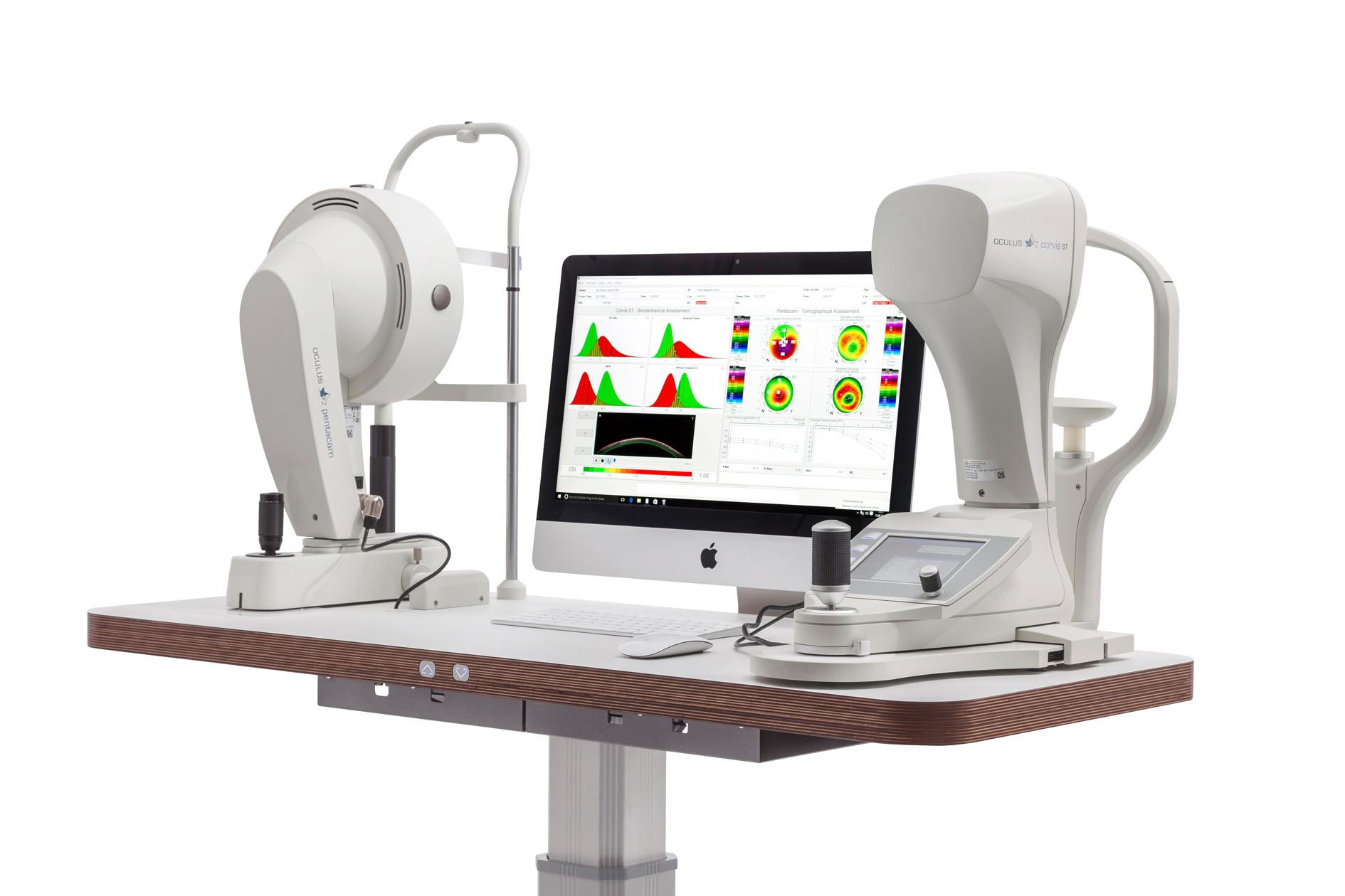 OCULUS Corvis (Corneal Biomechanics Test and Pachymetry)
It records the reaction of the cornea to a defined air pulse with a newly developed high-speed Scheimpflug camera that takes over 4,300 images per second. Intraocular pressure (IOP) and corneal thickness can be measured with great precision on the basis of these images. It studies the corneal biomechanics and reports the CBI index which helps in the detection of keratoconus. In addition, by integrating its data with that produced by the Pentacam, the TBI index is generated to optimize the detection of corneal ectasias.
 Pachymetry
It measures the thickness of all the corneal tissue, both central and peripheral.
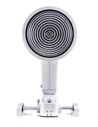 Keratograph 5M (K5M)
It allows to determine in a quantitative way the tear meniscus, tear break-up time and the analysis of the meibomian glands using infrared light. It is used for the diagnosis of dry eye.
 TearLab Osmolarity Test
An important technology for the diagnosis of Dry Eye Syndrome, it is an easy, simple, safe, fast and objective technique for measuring tear osmolarity (the degree of tear concentration).
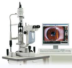 Digital Photography of the Anterior Segment
Using a special camera, photographs are taken of the anterior segment and adnexa as diagnostic images and to follow up on the evolution of different ocular pathologies. Digital Photography of the Posterior Segment
Using a special camera, photographs are taken of the inside of the eye in order to detect anomalies of the optic nerve, macula and other structures.
 Fluorescein Angiography
A contrast medium and a special camera are used to examine the blood flow in the retina and choroid. These are the two layers of the back of the eye.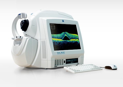 OCT CIRRUS 5000 (Optic Nerve and Retina)
A non-invasive diagnostic test that provides high resolution images of the retina and the optic nerve. It does not require contact with the eye because it is based on the reflection of light signals. It facilitates the diagnosis and management of retinal pathologies and glaucoma. OCT CIRRUS 6000
The most advanced model of Optical Coherence Tomography (OCT), designed for the detailed and precise diagnosis of various eye diseases. This device allows obtaining high-resolution and precise images of the internal structures of the eye, such as the retina and optic nerve. It is also used for studies of the anterior segment of the eye: cornea, anterior chamber and angle.
 OCTA (Optical Coherence Tomography Angiography)
Optical Coherence Tomography Angiography (OCTA) is a noninvasive imaging technique used to obtain detailed images of blood vessels in the retina and choroid of the eye. It provides precise information about the structure and blood flow in the ocular region.
 AcuTarget HD Analyzer
It uses double-pass technology to provide an objective measurement of vision quality. This tool is capable of quantifying the effects of ocular scatter on total vision. The diagnostic capabilities of this device can be used for the diagnosis of early cataracts, for the diagnosis of dry eye presenting without symptoms and for choosing the most appropriate refractive procedure for each patient.
 iCare HOME2 Tonometry
The iCare HOME2 measures intraocular pressure (IOP) for 24 hours. Unlike the measurement of intraocular pressure taken in the consultation office, the Ambulatory Pressure Monitoring allows obtaining a more complete and precise evaluation of changes in ocular pressure throughout the day.
-
 Exophthalmometry
It measures the degree of protrusion (forward displacement) of the eyeball.
 Binocular Vision Test
Used to determine whether both eyes are receiving visual stimuli at the same time. It also values stereoscopic vision which allows for the perception of depth, to capture what we see in three dimensions: length, width and depth.
The Schirmer test is used to evaluate the production of tears in the eyes. It is one of the diagnostic tools used to determine the type of dry eye and/or lacrimal gland dysfunction associated with autoimmune pathologies such as Sjögren's Syndrome.
The Titmus Test is a test to evaluate visual ability and depth perception in patients. Also known as a "stereoscopic vision test," the Titmus Test uses 3D images to determine visual acuity and binocular function.
It is also often used as a diagnostic aid for strabismus and amblyopia.
 Color Vision Test
Detects disorders of color perception.
 Contact Lens Fitting


